Witnessing endosymbiosis?
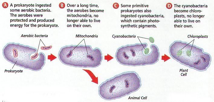
The most recent issue of Science has an intriguing report by a pair of Japanese scientists. They describe their discovery of a protist that appears to be in the early stages of an endosymbiotic process. For those of you who may be unfamiliar with the term, endosymbiosis refers to a specific association between two or more species: that one species lives inside the other one, the host, working together as one organism. The best examples we have of this involve engulfment of prokaryotes by eukaryotic cells: mitochondria in our own cells, and chloroplasts in plants.
(Continued below)
(Full-size picture can be found here)
Though there are several lines of evidence that support these ideas. Mitochodria and chloroplasts contain DNA, RNA, and ribosomes, all of which are similar to those of prokaryotes. Like free-living prokaryotes, mitochondria and chloroplasts divide by binary fission. Additionally, mitochodrion and chloroplasts each have two membranes. The outer one appears to be a product of membrane infolding by the host cell, but the inner one is probably the ancestral prokaryotes plasma membrane. Finally, DNA sequence comparisons have even identified the nearest modern relatives of the organelles: a species of rickettsia for mitochondria, and cyanobacteria for chloroplasts. However, there are always nay-sayers, who argue that because we haven't seen this happening, we don't have any good evidence that endosymbiosis happened with our ancestral cells.
In their current paper, Okamoto and Inouye state:
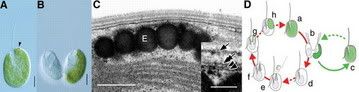
Fig. 1. (A) Hatena (ventral view). All green, symbiont-possessing cells have an eyespot at the cell apex (arrowhead). Scale bar, 10 µm. (B) A dividing cell (ventral view). The symbiont is always inherited by only one of the daughter cells. Scale bar, 10 µm. (C) The ultrastructure of eyespot integration (longitudinal view). E, eyespot granules. Scale bar, 400 nm. Inset: A magnified view of the membranes. The inner and outer plastid membranes (arrowheads), the single symbiont-enveloping membrane (double arrow), and the host plasma membrane (arrow) are tightly layered. Scale bar, 50 nm. (D) The life cycle of Hatena, based on observations of natural populations. Hatena alternates between a host phase that harbors a green endosymbiont and a predator phase that acquires the endosymbiont after division. Solid line, observed steps in the process; broken line, hypothetical steps.
It's not just simply an engulfment, either. The symbiont keeps some of its own organelles (such as its nucleus, mitochondria, and plastid), but loses others (such as its flagella and cytoskeleton). Additionally, the symbiont-derived eyespot (the arrow in figure 1A) seems to always be located in the same place within the host cell--at the juncture of four membranes derived from the symbiont, the symbiont's plastid, and the host. Finally, in experiments using symbiont-free hosts and a different strain of the Nephroselmis symbiont did not result in the phenotypic changes observed with the symbiont strain, suggesting specificity.
This isn't the first time a phenomenon like this has been observed in the lab. An endosymbiotic relationship between Amoeba proteus and a symbiont termed "X-bacteria" was first observed ~40 years ago. But of course, while biologists marvel at fascinating phenomena such as these, creationists can just sit back and spout, "but they're still amoeba!" What a boring life that must be.
(Continued below)
(Full-size picture can be found here)
Though there are several lines of evidence that support these ideas. Mitochodria and chloroplasts contain DNA, RNA, and ribosomes, all of which are similar to those of prokaryotes. Like free-living prokaryotes, mitochondria and chloroplasts divide by binary fission. Additionally, mitochodrion and chloroplasts each have two membranes. The outer one appears to be a product of membrane infolding by the host cell, but the inner one is probably the ancestral prokaryotes plasma membrane. Finally, DNA sequence comparisons have even identified the nearest modern relatives of the organelles: a species of rickettsia for mitochondria, and cyanobacteria for chloroplasts. However, there are always nay-sayers, who argue that because we haven't seen this happening, we don't have any good evidence that endosymbiosis happened with our ancestral cells.
In their current paper, Okamoto and Inouye state:
Here we describe a flagellate (Fig. 1A--see below) that appears to be in the formative stages of an ongoing endosymbiosis. The flagellate, which we tentatively refer to as Hatena ("enigmatic" in Japanese), will be formally described as a member of a recently elected division Katablepharidophyta. Hatena is currently uncultivable, so cells from natural populations were used for investigations. Nearly all the cells had a green "plastid" with an eyespot at the cell apex. However, this plastid was inherited by only one daughter cell (Fig. 1B), indicating the structure is a symbiont.
Fig. 1. (A) Hatena (ventral view). All green, symbiont-possessing cells have an eyespot at the cell apex (arrowhead). Scale bar, 10 µm. (B) A dividing cell (ventral view). The symbiont is always inherited by only one of the daughter cells. Scale bar, 10 µm. (C) The ultrastructure of eyespot integration (longitudinal view). E, eyespot granules. Scale bar, 400 nm. Inset: A magnified view of the membranes. The inner and outer plastid membranes (arrowheads), the single symbiont-enveloping membrane (double arrow), and the host plasma membrane (arrow) are tightly layered. Scale bar, 50 nm. (D) The life cycle of Hatena, based on observations of natural populations. Hatena alternates between a host phase that harbors a green endosymbiont and a predator phase that acquires the endosymbiont after division. Solid line, observed steps in the process; broken line, hypothetical steps.
It's not just simply an engulfment, either. The symbiont keeps some of its own organelles (such as its nucleus, mitochondria, and plastid), but loses others (such as its flagella and cytoskeleton). Additionally, the symbiont-derived eyespot (the arrow in figure 1A) seems to always be located in the same place within the host cell--at the juncture of four membranes derived from the symbiont, the symbiont's plastid, and the host. Finally, in experiments using symbiont-free hosts and a different strain of the Nephroselmis symbiont did not result in the phenotypic changes observed with the symbiont strain, suggesting specificity.
This isn't the first time a phenomenon like this has been observed in the lab. An endosymbiotic relationship between Amoeba proteus and a symbiont termed "X-bacteria" was first observed ~40 years ago. But of course, while biologists marvel at fascinating phenomena such as these, creationists can just sit back and spout, "but they're still amoeba!" What a boring life that must be.